Regular eye exams are integral to optimal eye health, as many eye conditions develop without noticeable symptoms or warning signs. During a standard exam, your optometrist will evaluate your vision and check for signs of eye diseases like macular degeneration, glaucoma, and other potential issues. Routine eye exams will also include tests to check various aspects of your eyes, such as colour vision and sharpness.
Contents
- 1 What Does a Standard Eye Exam Test For?
- 2 Introduction and Medical History
- 3 General Vision Test with the Snellen Chart
- 4 Visual Fields
- 5 Refraction Test
- 6 Ocular Motility Exam
- 7 Colour Test
- 8 Ophthalmoscopy
- 9 Eye Puff Test
- 10 Slit-Lamp Exam
- 11 How Often Should I Schedule a Standard Eye Exam?
- 12 What Should I Do After My Eye Exam?
What Does a Standard Eye Exam Test For?
A standard eye exam evaluates various aspects of your vision and eye health, including:
- Visual Acuity: How clearly you can see at different distances.
- Visual Field: Your peripheral (side) vision range.
- Signs of Eye Disease: Screening for glaucoma, cataracts, and macular degeneration.
- Eye Movement: How well your eyes track and move together.
- Colour Vision: Ability to distinguish between different colours.
- Eye Structure Condition: Assessing the overall health of your eyes, including the cornea, lens, and surrounding tissues.
- Retinal Health: Examining the retina for signs of damage or disease.
Introduction and Medical History
If you’re a new patient, the optometrist will ask you to complete paperwork about your medical history, personal information, and any symptoms or issues you have experienced regarding your vision. This information will give your optometrist a general understanding if you are at risk for any eye condition.
Be sure to include other relevant details, such as:
- Your current eyeglass or contact lens prescription
- Any pre-existing eye conditions
- Previous treatments or care provided by other eye specialists
Once your paperwork is complete, you can meet your eye doctor. They will take you into their office, review your information, and ask follow-up questions regarding any symptoms or vision problems impacting your sight.
General Vision Test with the Snellen Chart
You’ve probably experienced the Snellen Chart during a routine eye exam or check-up with your family doctor. This standard vision test uses a large chart of letters ranging in size. The doctor will ask you to place one hand over one eye and read out any letter they point at. Then, you’ll do the same with the other eye, which tests the sharpness of your vision.
Visual Acuity
The Snellen Chart is placed 20 feet away from you and shows letters in varying sizes to gauge how clear your vision is. The smallest letter you can read is your visual acuity.
- 20/20 Vision: This means you can see clearly at 20 feet what a person with normal vision should see at the same distance — considered perfect eyesight.
- 20/60 Vision: If you have 20/60 vision, you would see at 20 feet what someone with normal vision could see from 60 feet away, indicating reduced clarity.
Another tool to test your visual acuity is the occluder. The occluder is a tool that covers one eye at a time for your optometrist to test your visual acuity. Often, they ask you how many fingers they are holding up to verify your vision.
Visual Fields
The visual fields test measures your field of vision, including your peripheral (side) vision. It helps detect potential issues related to underlying conditions such as glaucoma, brain tumors, or other neurological disorders.
This test involves sitting at a machine called a perimeter. Your optometrist may ask you to sit up close to assess how far you can see in all directions. They may also use flashlights to detect any areas of vision loss.
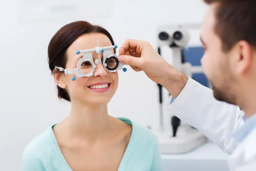
Refraction Test
A refraction test determines whether you have refractive errors in your vision. It assesses blurriness associated with conditions such as astigmatism, nearsightedness, and farsightedness.
Retinoscope
Your doctor may use a retinoscope or computerized vision-testing instrument by pointing a light at your eyes. That gives them an estimate of your prescription strength in correlation with your vision. As they monitor your vision, they’ll adjust the tool, often combined with a phoropter, by placing different lenses in front of your eyes to determine which is best for giving you clear, sharp sight.
Phoropter
This tool is a large metal frame with various lenses that the optometrist can switch to test which lens increases your vision. The optometrist adjusts the lens or changes the grade to determine the refraction rate and the patient’s required prescription.
Ocular Motility Exam
During this part of your eye exam, your optometrist will test your eyes’ movement, reflexes and tracking.
Cover Test
This simple test requires a tool to cover one of the patient’s eyes. The optometrist then assesses the movement of the other eye. The test involves keeping your eye on an object, like a pen, and looking in different directions without moving your head. This will assess any vision restrictions or weaknesses in your eye movements, determining if either eye is unaligned, whether you have lazy eyes, are experiencing a loss of depth perception, or have crossed eyes.
Colour Test
Seeing the world in colour is a luxury many people don’t have due to a deficiency in their vision. This test will determine the scope of colour vision, which involves looking at cards with coloured dots that include numbers. The test will involve the optometrist asking whether you can see a specific number. If you can, your colour vision is good, but if not, they may need to perform more tests to determine why.
Ophthalmoscopy
During this test, the optometrist uses an ophthalmoscope, a tool that functions like a mini telescope, to examine the external and internal structures of the eyes. They inspect the skin around the eyes, eyelids, eye surface, lens, and cornea. This test helps detect conditions such as retinal detachment, glaucoma, and systemic health issues like diabetes or high blood pressure, which can impact eye health.
Pupillary Tests
An optometrist will drop special eye drops into your eyes to encourage papillary dilation. This allows the optometrist to get a better look at the internal structure of the eyes. The effect of the drops can last for as much as twenty minutes and could cause some vision difficulties. Still, this test is critical for assessing underlying health and potential neurological problems.
Eye Puff Test
The eye puff test measures the pressure of the cornea. During this quick test, air will be “puffed” into both of your eyes so the doctor can analyze the fluid pressure inside them. If the eye pressure is higher than usual, you may be at risk of glaucoma.
Slit-Lamp Exam
To finish off the test, the doctor will use a slit lamp, also known as a biomicroscope. This device illuminates the eyes, allowing the optometrist to see the eye structure, the front and back of the eyes, and the eyelids.
During this exam, the optometrist can observe the lens behind your pupil. This zoomed-in scope allows them to assess for certain risks, such as diabetic retinopathy and cataracts, or whether you require more than a new glasses prescription.
How Often Should I Schedule a Standard Eye Exam?
Determining exactly how often you should have your eyes tested will vary depending on age, health conditions, family history, and whether or not you’re currently experiencing any issues with your vision. Typically, it should be every two years and annually for children. That’s why it’s always best to talk to your doctor so they can perform a preliminary test to determine how frequently you should be getting them tested.
What Should I Do After My Eye Exam?
Once the check-up is complete, your optometrist will discuss any required eye care, prescriptions, or follow-ups and walk you through their findings. They may also refer you to a specialist if any issues or conditions arise.
If it’s been longer than you can remember since you last checked your eyes, visit Laurier Optical on Innes or contact us today to arrange an eye exam.